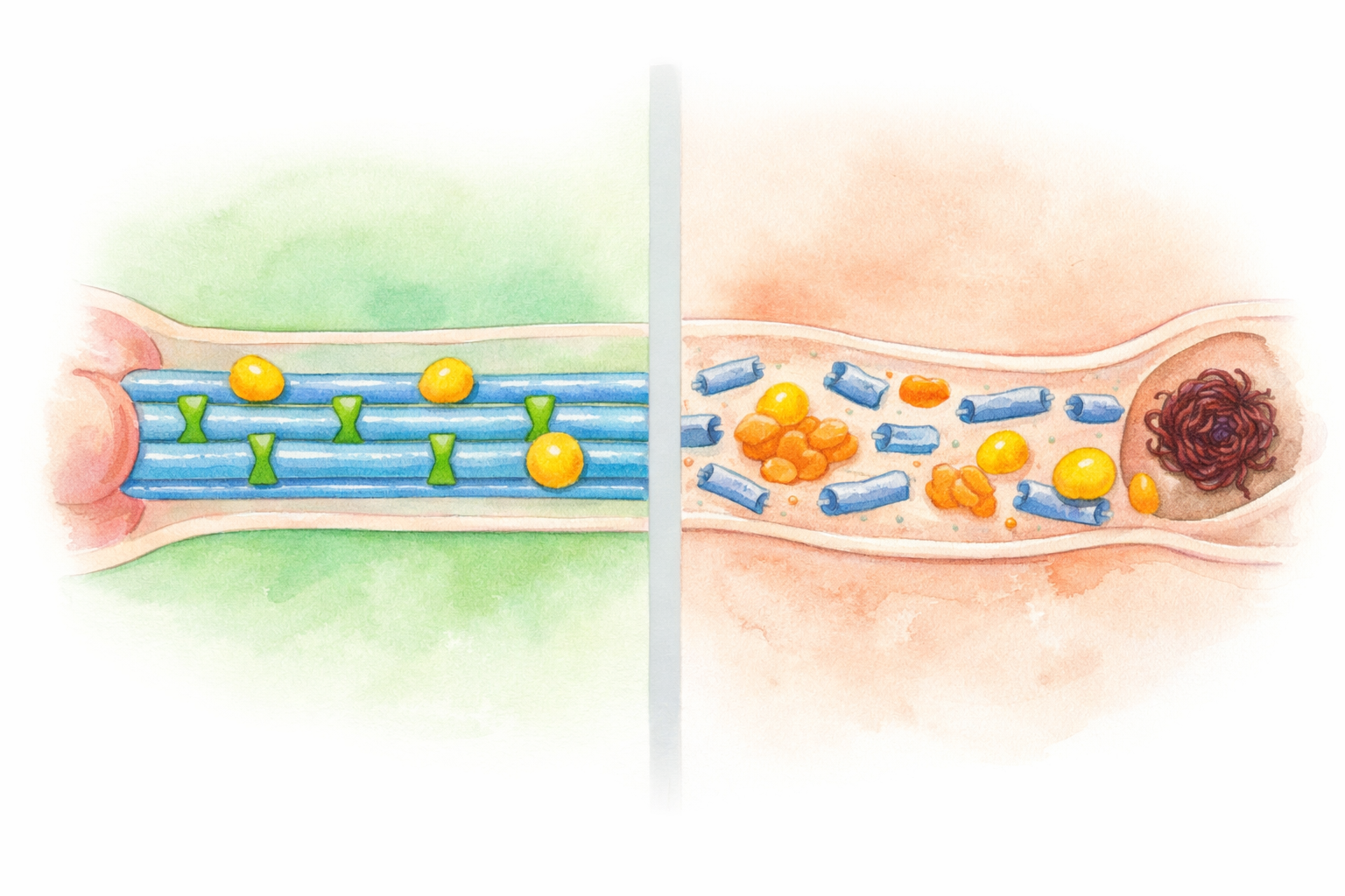
Side-by-side comparison of a healthy neuron axon with intact blue microtubules, green tau cross-ties, and yellow transport vesicles on the left, and a diseased neuron axon with fragmented microtubules, detached orange hyperphosphorylated tau, dark red neurofibrillary tangles, and stranded vesicles on the right
Hover over a numbered marker or a label to compare healthy and diseased tau.

Edit Mode — drag markers to calibrate positions

Drag a marker to see its live coordinates.